New dental patient exams by Advanced Dentistry of Richmond
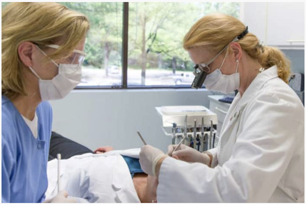
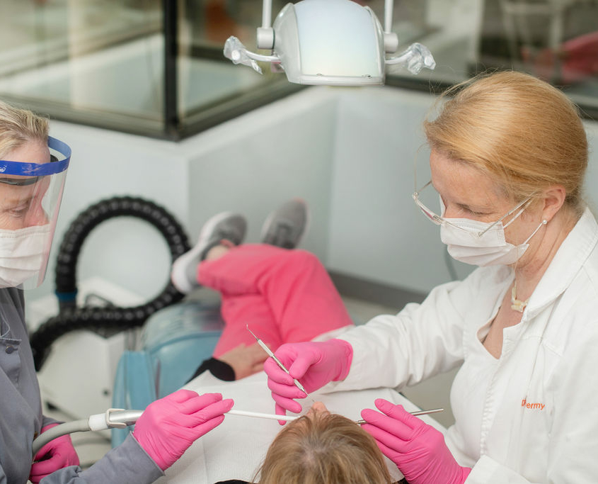
A clinical exam is more commonly referred to as a routine check-up. This check-up lets Dr.Klostermyer essentially take inventory of the overall health, of your temporal-mandibular joint TMJ (where your jaw attaches to your skull), and all of your teeth and diagnose any potential problems you might have.
First Dr.Klostermyer will visually check your face and neck for any abnormalities, such as lumps, bumps, or swellings. She will carefully examine your TMJ.
What do we do during our cancer screening?
After the extra-oral examination, she will check the inside of your mouth. During this cancer screening part of the check-up, she will be looking for any abnormalities in the soft tissue, such as discolorations or ulcers on your lips, gums, tongue, palate, and cheeks.
Our thorough check covers gums, jawbone, gingivitis, gum disease, and bone disease
Next Dr. Klostermyer will check your gums and jawbone, for any signs of gingivitis, gum disease, and bone disease. Then she will check your teeth for cavities and other problems like misalignment of the teeth, gum problems, and the presence or absence of wisdom teeth. She will be sure to look specifically at any areas where you may have symptoms or concerns.
New Patient Dental X-rays in Richmond Virginia
In most cases, a clinical exam by itself is not sufficient to completely diagnose all potential problems within your mouth. In fact, a majority of problems with your teeth and supporting bone and tissue are not visible to the naked eye. This is why x-rays play a key role in allowing a better, more accurate look. By using x-rays Dr.Klostermyer can check for any bone loss and determine the severity of any gum disease.
In addition to revealing any problems that were not visible during the clinical exam, these initial x-rays will also provide Dr.Klostermyer with a benchmark to reference during your future visits.
For everyone’s safety at Advanced Dentistry of Richmond, we use a state-of-the-art digital x-ray system to minimize exposure to potentially harmful x-rays.
Our Oral cancer screening
Every hour one American dies as a consequence of oral cancer! About 30,000 Americans are diagnosed with oral cancer each year. The disease has a higher mortality rate than cervical cancer, Hodgkin’s disease, liver cancer, or kidney cancer. Early detection is key. If caught early, a patient with oral cancer has a 90 percent cure rate. Who is at increased risk for contracting oral cancer? Those who:
- Use tobacco in any form
- Consume alcohol regularly combined with smoking
- Are you over the age of 40
- Have prolonged exposure to the sun (lip cancer)
Because 25 percent of the people diagnosed with oral cancer have no risk factors, an oral cancer screening should be a routine part of a regular dental check-up. This includes an examination of the entire mouth for the early detection of cancerous and pre-cancerous conditions.
Common symptoms of oral cancer or pre-cancerous cells include:
- A change in occlusion (bite)
- A color change in the oral tissues
- A tiny white or red spot or sore anywhere in the mouth
- Any sore that bleeds easily or refuses to heal
- Difficulty chewing, swallowing, speaking, or moving the jaw or tongue
- Lumps, thickenings, rough spots, or crusty areas
- Pain, tenderness, or numbness anywhere in the mouth, lips, or lymph nodes
Screening for oral cancer
During an oral cancer exam, Dr. Klostermyer will carefully examine your tongue, the inside of your mouth, and your lips to look for spots or sores that are flat, painless, white, or red. Many of these spots or sores are harmless, but some aren’t. So in some cases, a test may be needed to determine if a problem exists. This may involve a brush test, which involves collecting cells with a miniature brush from a suspicious sore or discolored area in the patient’s mouth. The cells will be sent to a lab for analysis. Should the results indicate symptomatic cells are present, a biopsy can then be performed.
Intraoral camera
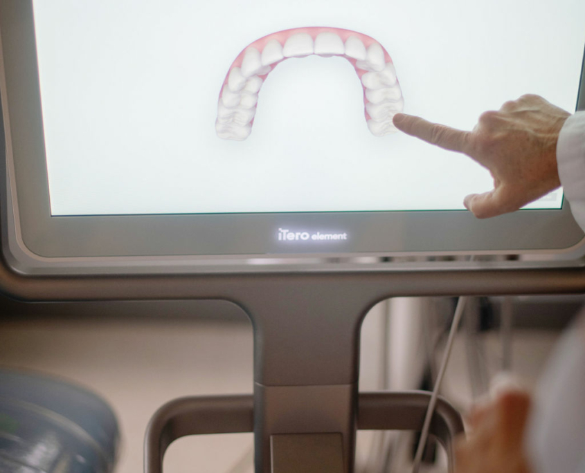
An intraoral camera is a lightweight handpiece that is designed for use inside the mouth. It uses LEDs for illumination and can be focused in any direction intra-orally to view and capture images and project them onto a computer screen.
Intraoral cameras provide detailed images which support an accurate diagnosis. Patients can also view the images recorded to better understand treatment plan options.

